MBI Videos
Robert Marc
-
 Robert Marc
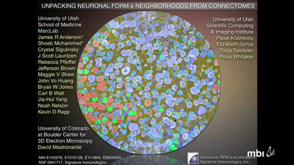
Mapping neural networks in brain, retina and spinal cord requires (1) comprehensive parts lists (vertex types), (2) nanometer scale connection detection (edge types), and (3) millimeter scale network tracing. Connectomics based on high-resolution automated transmission electron microscope imaging merges these operations and allows discovery of network modules and motifs as well as the their geometric patterning cell shapes.
The mammalian retina contains ≈ 70 classes of neurons assembled into ≈ 15 different network modules, with significant motif overlap. We analyzed mammalian retinal connectome RC1 to unpack this mesh of neurons. A key question that emerges in tracing networks is how neurons and patterns are regulated. I will describe the scales and modes of patterning (packing, tiling, covering) adopted by different neurons and discuss the implications for developmental regulation.
-
 Robert Marc
Mapping neural networks in brain, retina and spinal cord requires (1) comprehensive parts lists (vertex types), (2) nanometer scale connection detection (edge types), and (3) millimeter scale network tracing. Together this requires high resolution transmission electron microscope (TEM) imaging on a scale not routinely possible. Combining serial sectioning and TEM hardware control (SerialEM, Mastronarde, 2005, PMID 16182563) it is possible to created automated TEM (ATEM) imaging of mammalian retina composed of ≈0.4-1.4M high resolution images and assemble them into coherent 3D volumes of 16-21 TB of raw image data (Anderson et al., 2009, PMID 19855814). How should we build even larger connectomes? At present, we estimate that 100—1000 TEM systems are underutilized globally, representing the most cost-effective scale-up path.
The next ask is navigating, exploring, segmenting and annotating the data space for characterization of network motifs. The key tool for this is the Viking system developed by Anderson et al. (2010 PMID 21118201, 2011 PMID 21311605). Viking allows web-compliant delivery of imagery and collection of markup. Our experience suggests that complex network analysis (rather than simple shape-building) is more effectively done by teams of trained annotators working under executive analysts rather than crowd-sourcing. At present, retinal connectome RC1 contains >825,000 discrete annotations tracking ≈ 680 neural structures with 8300 connections, and ≈ 300 glia. We estimate this is only ≈20% of the expected annotation set. However, efficiency accelerates 2-3 fold as the density of identified process collision increases. Further many connection sets are replicates
Lacking robust tools for automated tracking, the best strategy has been to use multichannel molecular / activity markers and cell classification to segment and prioritize tracking targets, followed by intensive manual annotation (Lauritzen et al., 2012 PMID 23042441). A scientifically credible yet computationally manageable data volume in the vertebrate nervous system is currently in the 10-50 TB range. But a persistent challenge has been the vast scale differences for different neurons in a volume. There are an estimated 60+ classes of cells in the mammalian retina. For example retinal connectome RC1 is a disk of neural retina 243 μm wide and 30 μm tall. It contains 104 copies of the single class of rod bipolar cells with axonal fields spanning 15-25 μm; 39 copies of one of its target cells the AII amacrine cell, whose dendrites span 50-60 μm; and a dozen copies of a larger target, the AI amacrine cell with dendrites spanning up to 1000 μm. But one of the main targets of the is the ganglion cell superclass, which can be segmented into 15 classes. RC1 contains a few whole copies of a few of these, and fragments of many more, but a volume containing a complete set would need to be > 1 mm in diameter and require over 250 TB. While this is computationally feasible, the acquisition time for one microscope is not practical, nor is routine small-team annotation. Clearly, scaling by (1) platform and (2) automated annotation is critical.
Finally, some key networks in the retina are massive structured hubs. The AII amacrine cells has partnerships with at least 30 cell classes, meaning that all retinal neurons are no more than 2 hops from an AII cell and most no more than 2 hops from any other cell. This means that network graphs for even small volumes are extremely dense and differential visualization is a major goal.
